The foot and ankle are made up of 26 bones, 33 joints and more than 100 muscles, tendons, and ligaments. The anatomy of the foot is separated into three main parts: the forefoot, which is five metatarsal bones, and the toes (phalanges); the midfoot, which contains five tarsal bones — the navicular, cuboid, and three cuneiform that form the arch of the foot; and the hindfoot, which forms the heel (calcaneal) and is the largest tarsal bone, and the talus which forms the pivot of the ankle and connects to the bones of the leg.

The primary muscles of the foot include: the anterior tibial, which allows the foot to flex upward; the posterior tibial, which provides support to the arch; the peroneal tibial, which controls movement of the outer ankle; the extensors, which help the ankle raise the toes to begin the action of stepping forward; and the flexors, which stabilize the toes against the ground.
The muscles of the foot are connected to bones by elastic tissue called tendons. The largest, strongest and most important tendon of the foot is the Achilles tendon, which extends from the calf muscle to the heel.
Related Posts
 ‘High-Level’ Football Players Return to Play After ACL Reconstruction
 Stacey Margaret Jones: Reluctantly on the Disabled List (and What to Do About It)
 Weight of the Razorback World Rests on Brandon Allen’s Shoulder
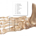 Study Shows Concussions Increase Injury Risk in Athletes
 Do I Have Whiplash?
 BJ Maack – Mechanism of Injury