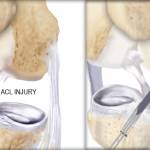
The knee is one of the most complex joints in the human body. There are four major bones that makeup the knee: the femur (thigh bone); tibia (shin bone); fibula (which runs parallel to the tibia); and the patella (knee cap).

What makes the knee so delicate is that when you bend your knee, it not only flexes and extends but slightly rotates as well. These movements are made possible by the vast array of muscles, including the hamstring, quadriceps, and ligaments that surround your knee joint to keep the bones in place.
Four major ligaments connect the femur to the tibia and are responsible for the controlled movement of your knee. These ligaments are: the medial collateral ligament; lateral collateral ligament; posterior cruciate ligament (PCL); and anterior cruciate ligament (ACL).
The ACL is one of the most important ligaments in the knee. It controls the movement of the lower leg by limiting the side-to-side rotation and preventing over-extension of the joint.
This major weight-bearing joint is cushioned by meniscal cartilage, which is a C-shaped tissue that is positioned in the joint between the tibia and the femur. Called the meniscus, it protects the joint and allows the bones to slide on each other free of friction and pain.
Related Posts
 ‘High-Level’ Football Players Return to Play After ACL Reconstruction
 Stacey Margaret Jones: Reluctantly on the Disabled List (and What to Do About It)
 Weight of the Razorback World Rests on Brandon Allen’s Shoulder
 Study Shows Concussions Increase Injury Risk in Athletes
 Do I Have Whiplash?
 BJ Maack – Mechanism of Injury